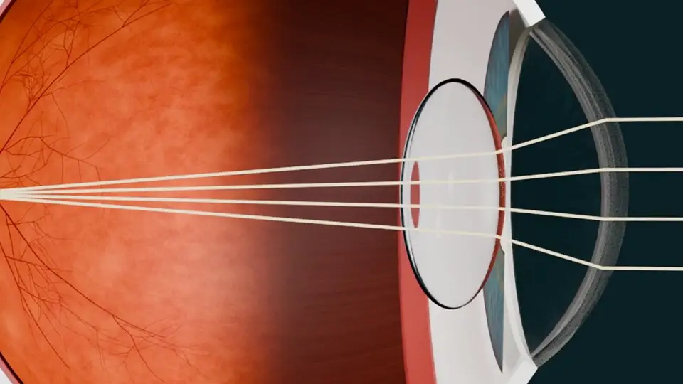
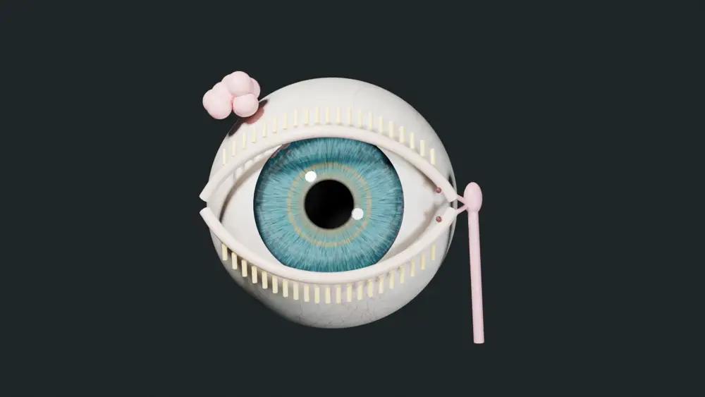
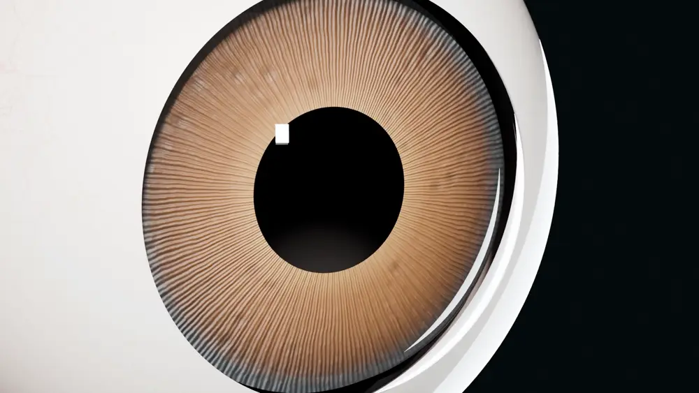
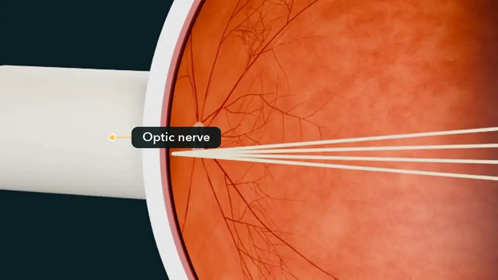
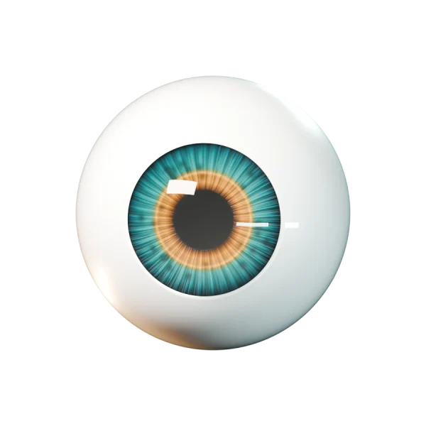
A few seconds from one of our first films, about the eye's lens.
Share how you explain things
You explain the same conditions, tests and treatments many times a week. Your best explanation can reach more patients.
1
Pick a topic
Any specialty. A condition, a test, a treatment, or what to expect after a procedure.
2
Record it
On your phone or computer, audio or video, 30 seconds to 4 minutes. Or upload one you recorded yourself, with no patient there.
3
Sign once
A short agreement in plain words: it's yours, it has no patient information, and we may turn it into films.
4
We make films
We check explanations against medical sources and turn clear ones into short patient films.
The one rule: explain it as you would to a patient, with no patient there. No recordings of real visits. No patient names, faces or details, and no charts or screens in view.
Credit, if you want it. "Based on an explanation by" your full name and credentials, your initials, or no name at all. You choose, and you can change it later.
The finished films, free for your patients. Films made from your explanations are yours to share with your patients at no cost.
Early access. You see new films and tools before they open to everyone.
You keep your recording. You give us a non-exclusive license, so you can still use it anywhere. You can ask us to stop future use at any time.
There's no payment unless we agree to one separately.
Our first films are about eye conditions
More specialties are next, starting with the topics doctors send us.
Retina
Blocked veins and arteries, retinal tears and detachment, floaters, macular hole and pucker, eye scans and more.
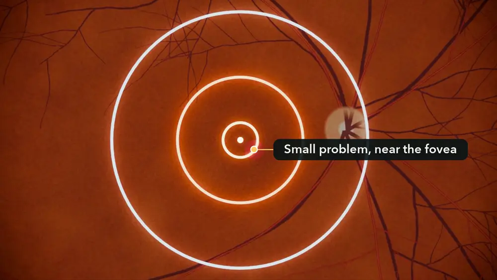
Still from: Central serous (CSC)
Eye Basics
Macular degeneration, diabetes and the eye, cataract, glaucoma, dry eye, pink eye, eyelids and more.
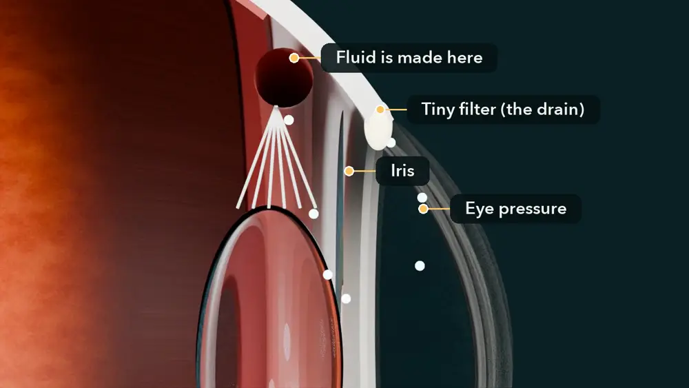
Still from: Glaucoma
Red eye: when to worry
The four warning signs that mean a red eye needs to be seen today.
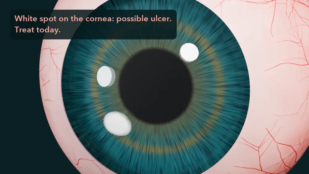
Still from: Red eye
How we check films for medical accuracy
Written from medical sources. Each script starts from published guidance and research on the topic.
Doctors are in charge. A retina specialist chooses and approves every eye-film topic. Explanations doctors send us will be the starting point for new films.
Films are checked. An automated reviewer checks the facts, the pictures and the words on screen, and problems it finds are fixed.
Fixes reach everyone. When we update a film, links already handed out play the new version.
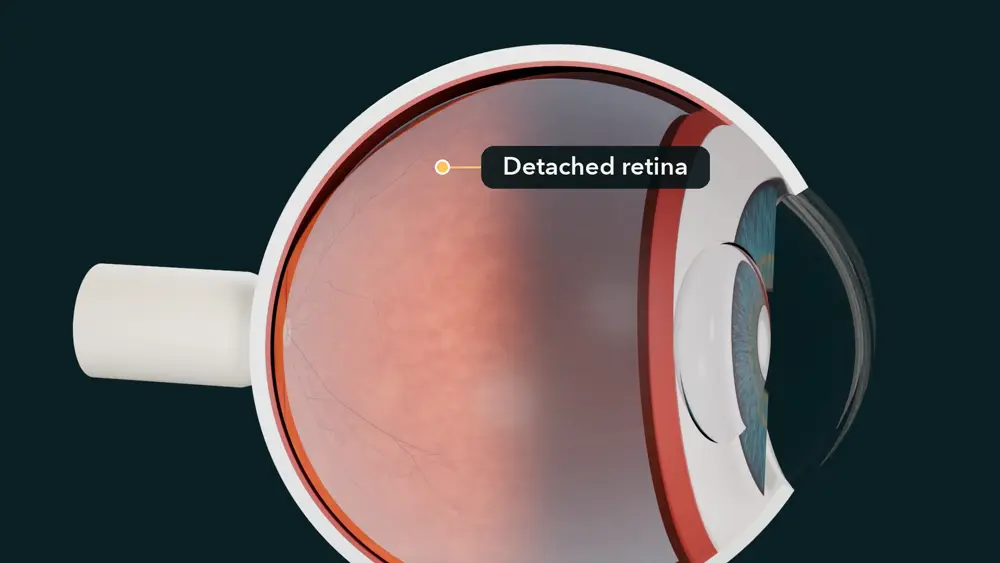
Still from our film on retinal detachment.
The films teach. They don't replace a doctor's advice or an exam, and they point patients back to their own doctor's plan.
What these films are
Short. About 2 minutes each.
Plain words. Models show what is happening inside the body. Captions are on screen.
Picked for you. Your doctor chose them for what was found at your visit.
Made with doctors. Our first films are about the eye, and an eye doctor chooses and approves every topic. The films are made with AI tools and checked before they go out. If you hear your doctor's voice, it may be an AI copy, made with that doctor's written consent.
Still from our film on cataract.
How to watch
1
Scan the code
Point your phone camera at the QR code from the office and tap the link. No app to install.
2
Watch while you wait
In the waiting room or the exam chair. Turn the sound down and read the captions, or use earbuds.
3
Or watch at home
Watch again later, or with family. The link works for 24 hours, or up to a week if your doctor chose that.
When the link ends, just ask the office for a new one.
Your privacy
No sign-in, and we never ask for your name.
The QR code holds only a list of films, a visit code and an end time. It has no name or health record. Anyone with the link can watch the films, so share it only with people you choose.
If you tap, type or say a question, it goes to your doctor's office team. So does a note that the link was opened. Both are deleted a few days after the link ends. Don't include your name or other private details. For anything urgent, call the office.
Some films play through YouTube, and YouTube's own privacy rules apply while they play. The others play from our own website.
Doctors who record explanations for us agree not to include any patient names, faces or details.
Still from our film on retinal detachment.
Questions?
The films help you understand. They don't replace your doctor. Write your questions down and talk to your doctor at your visit. If your vision or health changes suddenly, call your doctor right away. If you can't reach them, go to an emergency room.
The idea
Patients forget
Patients forget 40–80% of what they are told in a visit almost at once (Kessels, 2003).
Good explanations stay in the room
Every day, doctors explain conditions, tests and treatments well. Those explanations are heard once and then lost.
We keep them
We gather how real doctors explain medicine, with their consent and credit, check it, and turn it into patient films.
What we're building
A checked library of how real doctors explain medicine, across specialties: recordings, transcripts and the films made from them.
Content and data first. Our focus now is gathering explanations and making films, not selling to practices.
Consent built in. Every recording comes with a signed contributor agreement, is checked for patient information before use, and is credited the way its doctor chose.
Every specialty. Our first films are about eye conditions; more specialties are next.
Still from our Eye Basics film on glaucoma.
What works today
Live at teachme-eyes.web.app: a doctor taps the diagnoses and shows a QR code, and the films play on the patient's own phone. Links end after 24 hours by default, up to 1 week.
Our own clay-style eye films: a Retina series, Eye Basics, and more.
Doctors can record or upload explanations at /contribute/.
$0.35Cloud cost of one 2-minute film (measured; render and voice only)
From our own cost log, October 2026. It does not count the AI script or the doctor's review. These are facts about today, not promises of results.
Work with us
We'd like to hear from medical societies, schools, hospitals, health systems and investors who care about how patients learn.
 TeachMeMedical